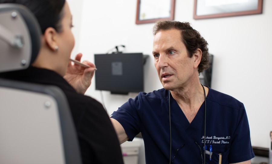
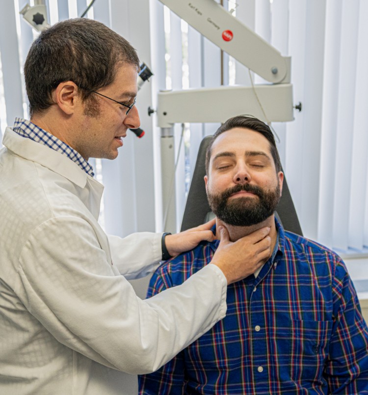
Swallowing Treatment

At The Voice and Swallowing Center, a division of ENT & Allergy Associates, LLP, is dedicated to the evaluation and treatment of voice and swallowing disorders and to further the understanding of voice and swallowing through education. The faculty at the Voice & Swallowing Center has invented or pioneered a suite of office-based diagnosis & therapeutic procedures.

Locate a Voice and Swallowing SpecialistLocate Speech Language Pathologist

Swallowing Treatment, or swallowing therapy, is divided into 4 broad categories: Behavioral Therapy, Dietary Therapy, Pharmacotherapy and Surgery. The purpose of swallowing therapy is to assure a safe swallow. By safe swallow, it is meant a swallow where the chance of choking is very small. Please click to the left to read more about the different types of swallowing therapy provided by the Voice & Swallowing Center.

Behavioral Therapy
Behavioral Therapy are postural maneuvers that are implemented to insure a safe swallow.

For example, a HEAD TURN is implemented when it is found that a patient is numb on one side of their throat during a FEESST exam. By having the patient turn their head to the numb side, the area of the throat that is numb becomes narrowed so that incoming food gets directed towards the sensate side of the throat.

Types of Swallowing Behavioral Therapy include:

1. Head Turn
By rotating the head to the side of either motor or sensory weakness, the patient can eliminate the injured side of the pharynx from the food bolus path. For an Endoscopic view of the effects of a head turn, please click here.

2. Chin Tuck
By placing the chin downwards toward the chest the patient:

  1. widens the vallecula to prevent bolus from entering airway
  2. puts the epiglottis in a more protective position
  3. narrows the laryngeal entrance

3. Effortfull Swallow
The patient is instructed to bear down, or to squeeze hard with all of their head and neck muscles while swallowing.

4. Shaker Exercises
Another way to open the upper esophageal sphincter by having the patient lay flat on their back and, in a precise manner, slowly lift their chin to their chest.

5. Supraglottic Swallow
4 step maneuver:

  1. inhale and hold breath (this closes the vocal folds)
  2. place bolus in swallow position
  3. swallow while holding breath
  4. cough after swallowing before inhaling (this clears any residue that may have entered the larynx)

6. Mendelsohn Maneuver
A technique that opens the upper esophageal sphincter. The patient is instructed to hold the thyroid cartilage up for several seconds. In this way, the larynx is kept tilted forward and elevated, thereby allowing the upper esophageal sphincter to relax.

Dietary Therapy
During the FEESST test it may become evident that certain types of food cause the patient to cough or choke, but other foods do not. During the FEESST exam the various food volumes and consistencies are used until it is determined which combinations allow the patient to swallow easily and safely.

Pharmacotherapy
Pharmacotherapy is therapy when certain medications may be prescribed which can help the patient swallow safely.

Depending on the results of the swallowing evaluation, certain medications may be prescribed which can help the patient swallow safely.

A) Mucolytic agents: Medications that thin-out thick secretions. Sometimes patients have very thick phlegm and mucus that makes it difficult for patients to swallow. Mucolytics can thin thick secretions so that they could be more readily expectorated and coughed.

B) Anti-acid medications: Swallowing problems are sometimes due to untreated, or insufficiently treated, acid reflux disease. The acid causes swelling in the throat which can contribute to swallowing difficulties. Under those circumstances, anti-acid medication is prescribed in order to help alleviate the throat swelling that may be contributing to the swallowing problem.

Surgery
The surgical management of the patient with dysphagia primarily depends on the etiology of the dysphagia. The more common etiologies of dysphagia that lend themselves to surgical correction are described.

A) Zenker's Diverticulectomy
Three procedures have been described to treat a Zenker's diverticulum: diverticulectomy, diverticulopexy, and peroral endoscopic division of the party wall between the diverticulum and the esophagus.

Diverticulectomy is usually selected for treating large diverticula in otherwise healthy patients (1). It involves an open-neck operation where the(Zenker's) hernia sac is identified and isolated. The sac is then resected its neck, taking care not to compromise the esophageal lumen by resecting too much mucosa, and closing the pharyngotomy with a watertight closure. A cricopharyngeal myotomy is performed as close to the posterior midline as is possible to minimize risk to the recurrent laryngeal nerves; the myotomy consists of dividing the entire circular cricopharyngeus muscle.

Diverticuloplexy, combined with cricopharyngeal myotomy, is preferred by some surgeons for dealing with small diverticula or large diverticula in high-risk patients. The sac is isolated and tacked with permanent suture to the prevertebral fascia, such that the mouth of the sac is in a dependent position (1). Diverticulopexy avoids a pharyngotomy, reducing the risk of a pharyngocutaneous fistula or injury to the recurrent laryngeal nerves.

Endoscopic peroral division of the party wall between the sac and the esophagus was first described by Dohlman in 1960 (2) . He used a special double-lipped esophagoscope, inserting one lip into the sac and one lip into the esophagus. Electrocautery was used to divide the party wall, including the cricopharyngeus muscle. Dohlman's procedure fell into disfavor because of an unacceptably high complication rate and mortality from mediastinitis. More recently, with some modifications to Dohlman's original technique such as utilization of an operating microscope and a laser, the endoscopic approach has gained acceptance, especially for very ill patients in whom an open procedure might pose greater risks.

B) Cricopharyngeal Myotomy
Dysphagia as a result of abnormalities with the cricopharyngeus muscle may be ameliorated by selective use of cricopharyngeal myotomy. Cricopharyngeal myotomy may be either surgical or pharmacologic (botulinum toxin). In general, cricopharyngeal myotomy is primarily useful for true cricopharyngeal achalasia such as after vagus nerve injury at the base of the skull where pharyngeal motor function remains otherwise intact (4, 5). Cricopharyngeal myotomy is contraindicated in conditions when there is impaired pharyngeal peristalsis or when significant reflux disease exists. Many disease entities where cricopharyngeal myotomy was thought to be useful in improving dysphagia, such as myopathy and brainstem stroke, may actually be of no benefit (6, 7 , 8).

C) Salivary Diversion Procedures
Dysphagia severe enough to result in the threat or actual circumstance of food and saliva constantly soiling the airway typically requires aggressive management. Surgical procedures that divert or diminish the flow of food and saliva from the airway include vocal fold medialization, tracheostomy, laryngeal stents, reversible laryngeal closure procedures, laryngotracheal separation and total laryngectomy. The application of any one of these treatment modalities depends on several patient factors such as underlying disease process and overall health status of the patient.

Patients who are aspirating regularly frequently become malnourished, which only exacerbates their underlying condition. Therefore, as measures are considered to prevent aspiration, alimentation through non-oral means should be implemented as well. Feeding gastrostomy or jejunostomy tubes, placed endoscopically (percutaneous endoscopic gastrostomy (PEG) or percutaneous endoscopic jejunostomy (PEJ)), are excellent ways to aliment patients who are at high risk for aspiration as a result of severe dysphagia.